Research

The focus of the research is on the maturation and adaptation of red blood cells (RBC).

After exiting the bone marrow, reticulocytes mature to form the highly adapted red blood cells travel through our circulation during their entire lifetime of in average 120 days. Thus, they are in constant move and adapt to their surrounding by shape changes, be it during in high-speed flow or during severe volume adaptations when they squeeze through small capillaries or the slits of the spleen having less than half their own size. While on the move, RBCs have to deal with continuous changes in oxygen tension and pH, have to scavenge reactive oxygen species and need to balance their responses towards the chemical and mechanical challenges.

In contrast, most of the current knowledge about RBCs as well as the diagnostic methods rely on RBCs in relative stasis, such as flux measurements, conventional patch-clamp, calorimetric assays, density centrifugation, atomic force microscopy. In extreme examples, the cells of investigation are even dead like in blood smears, electron microscopy or cyto-spins. Even if cells are on the move like in flow cytometers, they may rest in a drop of liquid. When taken from the circulation, the flow of the RBCs is suddenly absent and (together with the application of anticoagulants) the the RBCs experience a completely different environment that is likely to impair their properties.

The objective of EVIDENCE is the exploration of the properties and behaviour of RBCs under flow conditions and in vivo to understand pathophysiology and to design novel diagnostic devices. Theoretical models will help to understand these RBC properties and will enable the transfer of the gained knowledge into diagnostic devises in general and into the development of a spleen-on-the-chip in particular. Furthermore, we aim to understand the effect of the flow in bioreactors, allowing the efficient production of RBCs in vitro with the goal to produce RBC for transfusion.

The research programme is structured into five work packages (WP1 - WP5) that investigate specific topics:

  • WP1: In vivo investigation of RBC in circulation
  • WP2: Mathematical modelling of RBC in flow
  • WP3: Constructing a spleen-on-chip
  • WP4: Novel diagnostics for RBC diseases
  • WP5: Bioreactor concepts to upscale in vitro RBC production
     

WP1

In vivo investigation of RBC in circulation

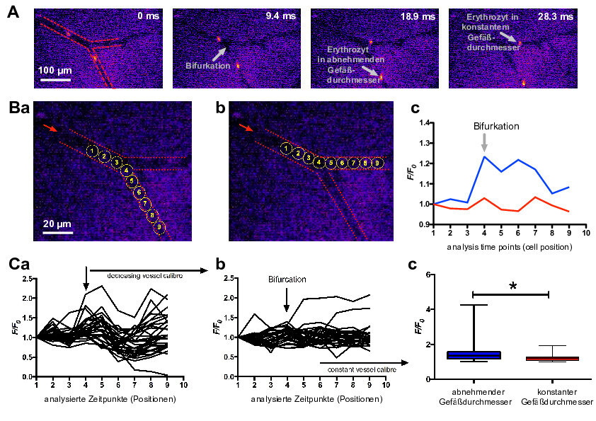
It is crucial to understand the properties and behaviour of RBC in vivo. In this work package we will study the shape of RBC that is vastly different between stasis and flow. However, to capture RBC in vivo in flow represents a technical challenge in itself, due to image resolution and image speed (frame rate) required to study these properties.

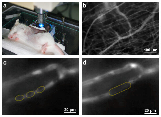
In vivo experiments will be performed by trans-illumination and fluorescence microscopy of mesenteric micro vessels and skin capillaries. This approach limits us to small rodents using a dorsal skinfold chamber. Measurements include single cell captures and the microvascular network. Initially, we will explore the general properties of blood cells, like RBC margination properties but we will also consider the exclusion of spleen filter function (splenectomised mice). 

Furthermore, we aim to understand pathophysiological situation. Therefore, we will investigate mouse transgenic disease models, like the Hbbth-4/Hbb+ mouse containing the human Β globin gene with the Β-thalassemia associated mutation, IVS-2-654 or the sickle cell anaemia Berkeley model.

To verify in vivo observations, we will perform complementary experiments in vitro using flow in microfluidic systems and the same pathophysiological RBCs as in the in vivo experiments. For the in vitro studies, we will use a recently developed 3D confocal microscopy approach that enables fast measurements4. For analysis, CTC will provide an automatic cell detection algorithm to analyse the flow based on real-time video recordings. This will include a controlled loop and cell shape analysis with a self-developed neural network based algorithm.

WP1

WP2

Theoretical modelling of blood cells in flow

From the mathematical modelling point of view, blood is a suspension of flexible objects, mostly RBCs, which are suspended in a complex solution - the blood plasma. Rich dynamics in oscillatory and pulsating flows occur at the scale of single cells at low haematocrit, which is similar to the smallest vascular capillaries.

Flow affects the shape of flexible objects, leading to cycling and chaotic dynamics. How microscopic dynamics couple with the macroscopic flow and if they can trigger flow instabilities themselves, are questions that we approach in collaboration between CNRS2 and USAAR2 within this work package.

Furthermore, in microvascular flow RBCs migrate to the centre of the flow, which results in a cell-free layer close to the boundary. This leads to a non-monotonic dependency of the flow resistance against tube diameter curve, known as the Fåhræus-Lindqvist-Effect. The white blood cells, which are a minority compared to the RBCs, are less deformable and migrate toward the boundaries, where they interact with the endothelium8. This leads to non-homogeneous concentration fields. These structure-forming mechanisms have been mostly studied in relation to simple channel flow. Therefore, a strong collaboration between WP1 and WP2 will provide in vivo observations to set the foundation for more sophisticated mathematical models.
 

Another element of complexity arises in blood flow, because the plasma macromolecules cause the RBCs to aggregate. At increased shear rates, these aggregates reversibly break up, which is the main mechanism of the pronounced shear thinning of blood. Both the concentration of the cells and the shear thinning character of the suspension are expected to significantly alter the flow instability mechanisms. Therefore, within EVIDENCE we perform direct comparisons between theoretical predictions and experiments on the flow of RBCs.

Experimental data is produced by means of microfluidic experiments in collaboration with INSERM, IBEC and RRM. We assess the flow of RBC suspensions and full blood in silico, in vitro and in vivo. We start at the level of single cells, investigating their shape as a function of the flow field by using microfluidic experiments combined with optical microscopy at diffraction-limited resolution. In the simulations, pathological cases are modelled by adapting the exact physicochemical parameters of the single RBCs to the parameters that have been obtained in the other WPs.

Having developed these experimental and computational methods, we will study the influence of pulsation on the flow and aggregation dynamics of the cells. We base simulations on the boundary integral methods, which enjoys high precision and the lattice Boltzmann method that is more rapid and adapts to complex geometries.
 

WP3

Constructing a spleen-on-chip

RBCs interact with various cell types and tissues. Crucial is the interaction of RBC with the spleen structure and with splenic macrophages that probe RBC integrity and viability. EVIDENCE combines knowledge on advanced fluidic devices with real-time imaging that are further developed to imitate and investigate splenic function.

Fresh human spleen material obtained from organ donors is available through Sanquin. SB1 developed a protocol to purify (95%) human red pulp macrophages from human spleen to characterize their gene expression profile (RNAseq) and their phenotypic characterisation (flow cytometry).

The endothelium from the spleen is rather different from endothelium in other organs. Within EVIDENCE, IBEC and SB1 are working together to create artificial spleens composed of the sophisticated microfluidics devices developed by IBEC9 in which the human spleen cells, including red pulp macrophages and endothelium will be cultured. This unique setup mimics the human spleen as closely as possible.
 

RBC clearance in the spleen is increased during pathological conditions such as autoimmune haemolytic anaemia, sickle cell disease and malaria infection. CD47 was identified by SB1 on RBC as a potential "eat me" signal that is recognized by the SIRPalpha receptor on spleen macrophages. The identification of signals that govern RBC clearance is by both the lack of knowledge on the RBC phenotype and an incomplete characterization of the red pulp macrophages in the human spleen that phagocytose the RBCs. Shedding of "eat me" signal containing small membrane vesicles from the RBC surface is a mechanism whereby RBC avoid clearance by macrophages. The regulation of vesiculation is compromised in many RBC disorders, but little is known about the mechanism. VHIR and IBEC will investigate vesicle formation in health and disease.

WP4

Diagnostics for red blood cell related diseases

Anaemia is the consequence of intrinsic defects of the haematopoietic system leading to low RBC production (erythropoietic defects) or of defects of the RBC leading to premature clearance and, hence, decreased cellular survival (haemolysis). The underlying cause of rare anaemias is unexplained in about 20% of patients. Anaemias of unknown origin (AUO) can be due to an erythroid defect, or to complex clinical situations and multifactorial mechanisms, in general associated with systemic, non-haematological, hereditary or acquired diseases.

The diagnosis of a rare anaemia is often prompted by pallor and fatigue and is often accompanied by jaundice. The proper diagnosis of a primary or secondary cause for anaemia and the identification of the defect in primary anaemia is a very important problem to solve for clinical and biological research. The technology driven SMEs will provide their know-how to develop new tests for RBC diagnosis together with experts in RBC biology (UZH, USAAR, INSERM, CRNS, Erytech). 

Erytech will use its RBC characterization toolbox (addressing the morphology, red blood cell rheology, eryptosis markers, micro particles release, immunogenicity, erythrophagocytosis as well as in-vivo bio distribution) in RBC-related diseases. Based on better understanding of the molecular mechanisms of pathology, new markers and approaches are identified and automated technologies for their detection developed.

Selected cells are screened by automated electrophysiology at Nanion for ion channel alterations as a possible cause of channelopathies. Especially the assays of the (automated) patch-clamp devices need to be tailored to specific RBC ion channels, as recently performed for Piezo1.
 

The electrophysiological approach is complemented by novel adhesion assays (INSERM), which allow the prediction of the thrombotic potential of RBCs. The combination of these technologies will allow more sophisticated and advanced diagnostic assays and tools, which are needed in the differential diagnosis of hereditary anaemias caused by:

  • membrane defects
  • enzyme defects
  • disorders of haemoglobin
  • disorders of RBC production

Technology will be tested taking advantage of transgenic mice models with altered membrane permeability (transient receptor potential channel knock-outs, e.g. TRPC6, or G-protein modification, e.g. Gq knock-out).

In line with these novel cell based approaches, EVIDENCE will develop a molecular screening tool for the diagnosis of hereditary anaemia based on Next Generation Sequencing (NGS) that has been proven successful in the identification of many genetic disorders. Based on NGS results, the aim of this work is a functional screening tool for the identification of genetic analysis of germline mutations. The development of this new diagnostic tool will allow for efficient integration of complete clinical data of patients and high-throughput data, resulting in faster, cost effective and reliable anaemia diagnosis.
 

WP4

WP5

Bioreactor concepts to upscale in vitro red blood cell production

Transfusion of RBC is associated with a risk for blood born disease and alloimmunisation, particularly upon recurrent transfusion for chronic anaemia. Cultured RBC, derived from stem cells or cell lines that lack blood group antigens, or enhanced with therapeutic proteins will enable precision medicine.

A single donor derived standard transfusion unit contains 1012 RBC of different ages and this number is the objective for large-scale cultures in novel types of bioreactors. SB2 and UB are involved in many aspects of RBC biosynthesis in bioreactors. The EVIDENCE network allows SB2 and UB to develop novel solutions for some of the challenges in the large-scale production of RBC. The overarching aim of this work package is to modulate RBC development program to optimize manufacture of RBCs for use in transfusion. The current culture process is expensive due to the reliance on commercial growth factors and large media volumes that are far from mimicking the bone marrow.

The first aim of this work package is to reduce costs and increase expansion efficiency of erythroid cultures by testing stable growth factor mimetics, and membrane bound growth factors in different types of bioreactor conditions. We will first focus on stem cell factor (SCF), because membrane bound and soluble SCF have distinct signalling properties, and loss of membrane bound SCF is associated with neonatal anaemia. Haematopoietic progenitor and erythroblast proliferation require the synergistic action of growth factor receptors, which reside in the membranes associated signalosomes. Signalosome formation changes during erythroid differentiation, but the process is ill defined. SB2 uses nitrogen cavitation for detergent-free isolation of small membrane fragments that are subsequently identified by mass spectroscopy. UB and SB will explore the effects of static and flow of growth factors on proliferation and signalling to upscale in vitro RBC production.

Erytech will characterize cultured erythrocytes (morphology, RBC rheology, eryptosis markers, micro particles release, immunogenicity, erythrophagocytosis as well as in vivo biodistribution) and compare them with native RBCs. We aim to identify critical parameters for in vitro RBC culture and behaviour to ensure the safety, improve the quality and the shelf life of RBCs as a transfusion product.

The second aim is to investigate the physical, cellular, and molecular effects of shear stress under distinct bioreactor conditions. It is unknown whether cell density or shear stress in these bioreactor culture conditions affect mechanosensing ion channels, Ca2+ signalling and proliferation. Both UB and SB have extensive proteomic data during expansion and differentiation. Those data are mined for ion channel expression and ion channel phosphorylation states will be compared cells at different stages grown in both static, stirring and wave bioreactors. CNRS1 and Nanion to measure respective ion channel activity.

The third aim is to increase our understanding of in vitro reticulocyte maturation, which is vital to improve the overall yield of blood production as reticulocyte filtration is less efficient than red cells with a ~50% yield after leukofiltration. SB2, UB, INSERM, USAAR1, CNRS1 and Erytech will study the additional changes, which occur during reticulocyte maturation events and compare this to disease states studied in WP4. We have shown that shear stress contributes to maturation but further studies are needed to explore this process further. UB, INSERM, and IBEC will use artificial spleens and other microfluidic systems to explore the cytoskeletal modifications that occur when retics are forced to alter shape and deform due to shear or constriction. Any alterations in kinase activity will be dissected using inhibitors and through CRISPR mediated modification of phospho